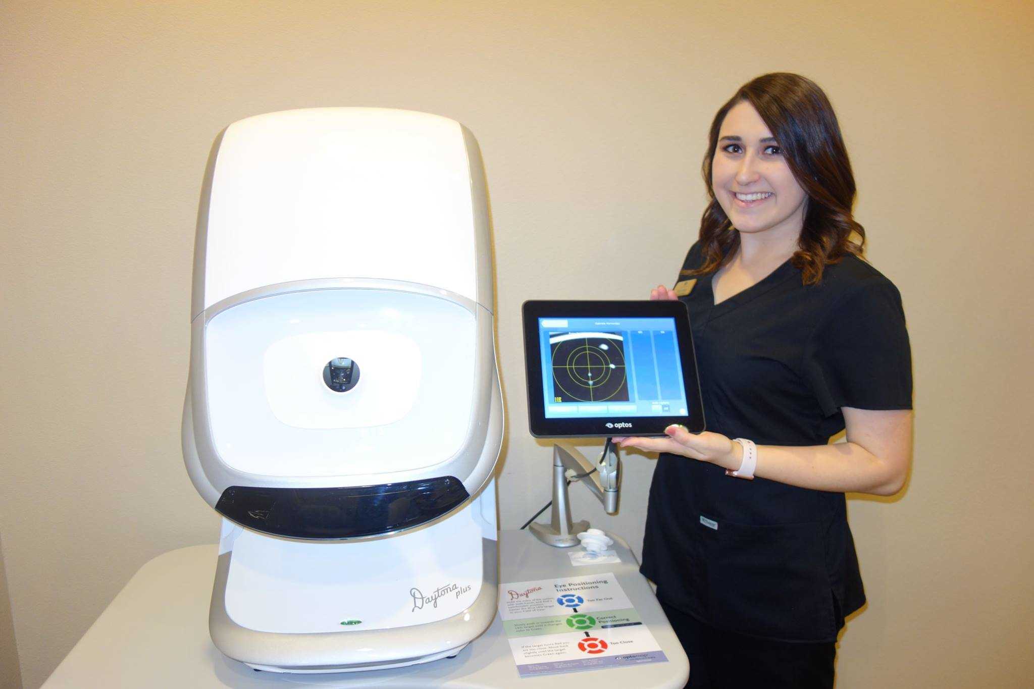
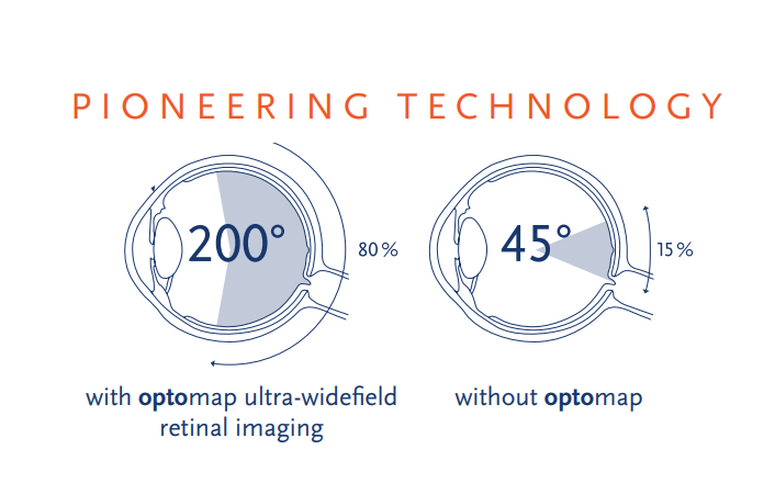
If your retina hurts, you want to check it with an Optos Daytona. It is a digital image scanner used to scan your retina. The ophthalmoscope’s scanning feature is a multi-laser that generates a 200-degree ultra-wide image of your retina.
An Optomap captures a retinal digital image equivalent to 80% of your retina. Its Virtual Scan Point technology captures images in a paltry 0.25 seconds. Your retinal image appears as a camera operator inside your eye took it.
Before its inception, retinal images captured by cameras could hardly exceed 45 degrees (15%). A typical camera could only capture the posterior pole of the retina. The size of the pupil needed cutting-edge technology that could see the peripheral arena.
Eye care specialists use this imaging technology to evaluate retinal disorders such as diabetic retinopathy and macular degeneration. It’s also capable of measuring freckles, known as choroidal nevus, at the back of the human eye. Developed by Optos, a company in Scotland, it has benefited 13 million users worldwide.
How does an Optos Daytona work?
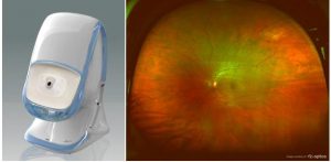
A Daytona Optomap uses a low-power multi-laser ophthalmoscope to capture digital scans of your retina. It employs the red and the green wavelengths to capture retinal images. It filters your image to evaluate several layers of your retina.
The Optomap comes with an ellipsoidal mirror that contains two focal points. The first focal point acts as a pathway for the laser light to focus on the retina, while the second focal point allows the patient’s eye to be positioned coincidentally.
The Optos Daytona greatly improves your image with a high resolution of 20 microns on-axis and 40 microns off-axis. This is better than its predecessor, the P-200, which had a 17-20-pixel resolution.
It also enhances retinal images with the two laser wavelengths that magnify and separate its different parts into color components, allowing for easier diagnosis.
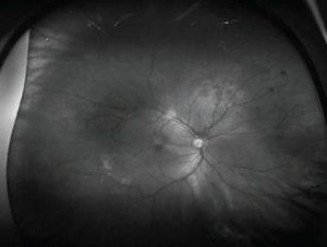
The two wavelengths, green and red lasers, work together to produce overlying green and red images, which appear pseudo-colored. The green laser penetrates the retina only to allow viewing of the retina and the anterior RPE, while the red laser enables viewing of the choroid and the posterior RPE.
The blue laser wavelength is also used for fluorescein angiograms. Unlike the green and red lasers, its usage is optional.
The value of the Daytona Optomap retinal imaging camera
The Daytona Optomap is a proven tool for maintaining your retinal health. It is not just a tool for the doctor; it allows you to participate in examining your eyes thoroughly. For professional eye specialists running medical services, you want to use Daytona.
-
Value to patients
Both children and adults find an Optomap useful because of its ease of use. Patients have a smooth experience using this high-end technology, and light cues in the Optomap guide patients in positioning themselves correctly.
Once in the right position, the Optomap automatically captures your retinal images with zero errors. Retinal imaging lasts only 2 minutes, and it takes actual pictures of your retina within a second.
Once captured, patients take another minute to get an explanation from their doctor. As expected, you’ll need to explain the images they are seeing to them. Explaining these images to your patients helps build their confidence in your service while presenting you as an expert in retinal disorder treatment.
This machine works seamlessly with an Electronic Health Record system. This means your patients’ retinal images will be transferred into their files. An EHR system also enables you to email your patients their retinal images for their personal records.
Before a Daytona Optomap examination, you must ensure your patients are aware of its use. This improves their experience with the Optomap Daytona and helps support your medical service through their referrals.
For eye specialists, training your staff on its use is essential for better medical services. Comprehensive training for using the Optomap instrument lasts 2 hours. This goes a long way toward making your medical services worthwhile.
-
Value to medical services
If you are offering a retinal medical service and intend to use the Optomap Daytona, there are vital factors you want to consider. You will need to do some math on your ROI –return on investment—how many patients you will need to break even, and whether you are making a full purchase or acquiring the instrument through leasing.
Return on investment
If you choose to purchase the Optos Daytona, it will cost you a substantial amount of dollars, up to $85000. However, your return on ROI is guaranteed as you are bound to make a $9000 profit.
You could make a profit of such magnitude with a few tweaks in the discharge of your services. You want to employ a pre-test stand that allows patients to take their retinal pictures and get an explanation from a doctor. This is achieved by investing in a pretest interest.
Your patient must understand every step of the process. Induct them through the technicalities of the process and the money she will need to spend. Ideally, this will portray somebody free to help and not just out for profit.
Red and Green Wavelengths: clinical relevance
The red and green wavelengths can penetrate different layers of your eyes to give different images, such as the red choroid and the green retina.
Choroidal melanoma
Choroidal melanoma is a common type of malignant tumor that implants itself in your eye. It stays undetected for an extended period as it rarely shows symptoms. On the contrary, patients who use an Optos Daytona can easily detect the condition.
Choroidal Nevus
The Choroidal Nevus arises from the accumulation of melanocytes in your choroid. These melanocytes occupy benign melanoma. Distinguishable characteristics of the choroidal nevi include a slate gray color and a slight elevation with distinct margins. They can only be seen in a red separation and not in green because of their location in the choroid.
Here are sample images of color melanoma and color nevus as captured by Daytona.
Color image of Melanoma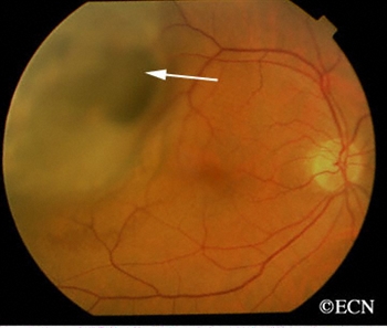
Color image of the Nevus
When you use a red wavelength, the images of Melanoma and Nevus appear dark. Please take a look at their pictures under a red wavelength.
Red melanoma
Red Nevus

Interestingly, the green wavelength produces slightly different images from the red. Have a look at the pictures of melanoma and nevus.
Green melanoma appears brighter than red melanoma.
Green Nevus appears darker when compared to the red Nevus.
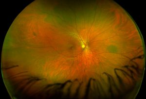
Chorioretinal atrophy
Chorioretinal atrophy is an unhealthy eye condition that occurs when the choroid and the retina wither away due to getting damaged. The outer layers forming the sensory retina, the RPE, and choriocapillaris all get damaged. It occurs when there is no circulation from the choriocapillaris.
Other symptoms accompanying Chorioretinal atrophy include pigment clumping within and around the lesion. This often arises from the migration of degraded RPE cells. These cells move into the inner layers of your retina.
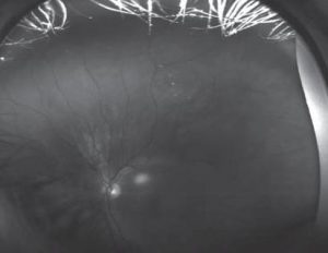
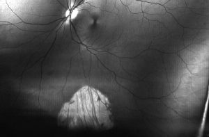
You can view the Daytona image of the Chorioretinal atrophy from different angles. You will find a choroid view at the posterior pole, while at the periphery, you will find a sclera that provides the lesion with a bright yellow color.
For a person with albinism or a very blonde fundus, a green laser penetrates the choroid to form a better choroid image than the image from a red laser. An image from a red laser gets scattered off the sclera. These Optomap Daytona images say it all.
Color image of a Chorioretinal atrophy
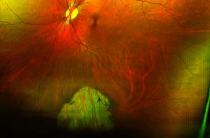
Red wavelength: choroid seen in both the RPE and the area of atrophy

Green wavelength: choroid is seen only through atrophy.
Advantages of the Optos Daytona
-
User-friendly
An Optos Daytona gives you an easy, short-time experience while guaranteeing quality. It comes with light cues that help you position yourself in the right spot for image capture. It works well with the elderly and children.
-
Wide field of view for Retina
Most conventional cameras can barely achieve a wide field of view. With Daytona technology, you get an ultra-wide view of your retina.
While conventional cameras capture images up to 45 degrees, the Optomap gives a wide field of view at 200 degrees. It also enables easy viewing of peripheral lesions that are difficult to see.
-
Wheelchair functionality
Daytona’s wheelchair functionality makes it elder-friendly. Older people with weak or bent limbs can easily use this machine, as it comes with a wheelchair, making it unnecessary to move a patient.
-
Excellent tool for education to patients and hospital staff
It is not as sophisticated a tool as you would imagine, but you can use it to induct staff on its usage and application to patients. Likewise, patients have an effortless experience trying to understand their complications.
For instance, patients are timelessly shown an Optomap of their eye while in the examination room. Suppose a diabetic patient sees a retinal hemorrhage. This has more impact on the patient than just being told to regulate your blood sugar.
-
Allows viewing through media opacities
The multi-laser ophthalmoscope penetrates the media better, giving a highly-resolved image. Compared to conventional fundus photography in cataracts, it provides a high-contrast image.
-
Images are clear
The Scanning laser ophthalmoscopy (SLO) produces exceptionally sharp images that give digital images a wide berth.
Disadvantages of the Optos Daytona
-
Image overexposure
Optomap images appear altered compared to DR’s fundus photography. Due to the deep focus of the Optomap, image structures that were previously invisible appear visible. You now have an image with floaters, eyelashes, lens opacities, and eyelids.
-
View distorted by Ellipsoid mirror.
The Ellipsoidal mirror isn’t the best choice for an Optomap. The fundus image appears elongated, and seeing things on the periphery is challenging.
-
Dark image
The image from Daytona appears darker than the traditional fundus camera. You want to solve this problem by increasing the illumination levels.
-
Small Pupils
For Optomap users, beware that it best works with pupils that are 3mm in diameter or more. If your pupil size is less than 2mm, pupil dilation may help enhance image quality.
-
Absent stereoscopic view
The stereoscopic view allows a monocular system. However, it loses information very quickly.
When should you use an Optomap?
For retinal checkups and examinations, a patient has to undergo a dilated BIO examination; even with an Optomap, a BIO examination can’t be eliminated.
An Optos Daytona works to complement the work of a dilated BIO examination. Only patients with specific critical conditions should use an Optos Daytona to examine their retinal problems. The Optomap imaging captures a wide range of conditions, including
- Retinal dystrophies
- Peripheral retinal degenerations
- Choroidal lesions & the
- Retinal vascular conditions
Conclusion
The digital image scanner Optos Daytona is here with us. It works best and surpasses other conventional cameras. For instance, it gives high-resolution images that can produce up to 200 degrees of an ultra-wide image.
The traditional camera does not have that magnitude of resolution and makes an image of up to 45 degrees. Eye care specialists use this tech-savvy tool to treat disorders such as diabetic retinopathy, macular degeneration, and hypertensive retinopathy.
If you run a medical service, you want to purchase this machine for your business as it is very user-friendly, and patients are bound to have an amazing experience. Optos Daytona retinal imaging is the best bet for people looking for retinal imaging and checkups.
Здесь вы найдете содержание 18+.
Контент подходит для взрослой аудитории.
У нас собраны разнообразные материалы.
Платформа предлагает четкие фото.
смотреть порно онлайн в хорошем качестве
Вход разрешен после подтверждения возраста.
Наслаждайтесь возможностью выбрать именно своё.
https://www.eater.com/users/roscartr
Здесь доступны учебные пособия для школьников.
Предоставляем материалы по всем основным предметам от математики до литературы.
Подготовьтесь к экзаменам с помощью тренажеров.
https://musecube.org/o-raznom/obrazovanie/gdz-po-informatike-k-uchebniku-bosovoj-dlja-8-klassov/
Демонстрационные варианты помогут разобраться с темой.
Доступ свободный для максимальной доступности.
Используйте ресурсы дома и успешно сдавайте экзамены.
На этом сайте вы найдете подготовительные ресурсы для учеников.
Курсы по ключевым дисциплинам включая естественные науки.
Успешно сдайте тесты с помощью тренажеров.
https://kgsxa.ru/components/com_newsfeeds/views/category/tmpl/news/3/1/4/359_kak_vibrat_horoshiy_sbornik.html
Примеры решений упростят процесс обучения.
Все материалы бесплатны для удобства обучения.
Используйте ресурсы дома и достигайте отличных результатов.
Модные образы для торжеств этого сезона вдохновляют дизайнеров.
Популярны пышные модели до колен из полупрозрачных тканей.
Детали из люрекса придают образу роскоши.
Многослойные юбки становятся хитами сезона.
Разрезы на юбках придают пикантности образу.
Ищите вдохновение в новых коллекциях — оригинальность и комфорт сделают ваш образ идеальным!
http://www.disneybounders.com/threads/%D0%9C%D0%BE%D0%B4%D0%BD%D1%8B%D0%B5-%D1%81%D0%B2%D0%B0%D0%B4%D0%B5%D0%B1%D0%BD%D1%8B%D0%B5-%D1%84%D0%B0%D1%81%D0%BE%D0%BD%D1%8B-%D1%8D%D1%82%D0%BE%D0%B3%D0%BE-%D0%B3%D0%BE%D0%B4%D0%B0-%E2%80%94-%D0%B3%D0%B4%D0%B5-%D0%BA%D1%83%D0%BF%D0%B8%D1%82%D1%8C.9048/
На данном сайте можно получить Telegram-бот “Глаз Бога”, что проверить всю информацию по человеку через открытые базы.
Бот функционирует по ФИО, обрабатывая доступные данные онлайн. Через бота доступны бесплатный поиск и полный отчет по запросу.
Сервис актуален на 2025 год и охватывает аудио-материалы. Бот сможет узнать данные в открытых базах и покажет информацию за секунды.
Глаз Бога glazboga.net
Это бот — выбор при поиске людей удаленно.
Здесь доступен Telegram-бот “Глаз Бога”, позволяющий проверить данные о гражданине через открытые базы.
Инструмент активно ищет по номеру телефона, обрабатывая публичные материалы в сети. С его помощью доступны пять пробивов и полный отчет по запросу.
Инструмент проверен на 2025 год и включает фото и видео. Сервис поможет проверить личность в соцсетях и предоставит сведения за секунды.
Глаз Бога
Это инструмент — выбор для проверки граждан через Telegram.
Здесь вы можете отыскать боту “Глаз Бога” , который позволяет проанализировать всю информацию о любом человеке из общедоступных баз .
Уникальный бот осуществляет проверку ФИО и предоставляет детали из онлайн-платформ.
С его помощью можно узнать контакты через Telegram-бот , используя фотографию в качестве начальных данных .
probiv-bot.pro
Система “Глаз Бога” автоматически анализирует информацию из проверенных ресурсов, формируя исчерпывающий результат.
Клиенты бота получают ограниченное тестирование для ознакомления с функционалом .
Платформа постоянно совершенствуется , сохраняя актуальность данных в соответствии с требованиями времени .
На данном сайте доступен мессенджер-бот “Глаз Бога”, что проверить всю информацию о человеке из открытых источников.
Инструмент функционирует по номеру телефона, используя публичные материалы в сети. Благодаря ему осуществляется бесплатный поиск и глубокий сбор по запросу.
Сервис проверен на 2025 год и поддерживает мультимедийные данные. Глаз Бога поможет найти профили в соцсетях и покажет информацию мгновенно.
https://glazboga.net/
Данный сервис — идеальное решение при поиске персон онлайн.
Здесь вы можете получить доступ к боту “Глаз Бога” , который способен проанализировать всю информацию о любом человеке из общедоступных баз .
Данный сервис осуществляет проверку ФИО и показывает информацию из соцсетей .
С его помощью можно узнать контакты через официальный сервис , используя имя и фамилию в качестве ключевого параметра.
пробить номер
Алгоритм “Глаз Бога” автоматически собирает информацию из проверенных ресурсов, формируя структурированные данные .
Подписчики бота получают ограниченное тестирование для тестирования возможностей .
Платформа постоянно обновляется , сохраняя высокую точность в соответствии с требованиями времени .
На данном сайте можно получить сервис “Глаз Бога”, что проверить всю информацию о человеке по публичным данным.
Сервис функционирует по ФИО, обрабатывая актуальные базы в Рунете. Через бота осуществляется пять пробивов и глубокий сбор по имени.
Сервис обновлен на 2025 год и включает мультимедийные данные. Глаз Бога гарантирует узнать данные в открытых базах и покажет информацию в режиме реального времени.
https://glazboga.net/
Такой бот — идеальное решение при поиске граждан удаленно.
В этом ресурсе вы можете получить доступ к боту “Глаз Бога” , который способен собрать всю информацию о любом человеке из общедоступных баз .
Этот мощный инструмент осуществляет проверку ФИО и предоставляет детали из государственных реестров .
С его помощью можно проверить личность через Telegram-бот , используя автомобильный номер в качестве ключевого параметра.
проверка номера телефона
Система “Глаз Бога” автоматически анализирует информацию из открытых баз , формируя структурированные данные .
Пользователи бота получают пробный доступ для проверки эффективности.
Платформа постоянно развивается, сохраняя скорость обработки в соответствии с требованиями времени .
Прямо здесь доступен Telegram-бот “Глаз Бога”, который собрать сведения по человеку по публичным данным.
Инструмент активно ищет по номеру телефона, анализируя актуальные базы в Рунете. Благодаря ему можно получить 5 бесплатных проверок и глубокий сбор по фото.
Платформа проверен согласно последним данным и включает мультимедийные данные. Глаз Бога гарантирует узнать данные по госреестрам и отобразит результаты мгновенно.
https://glazboga.net/
Данный бот — идеальное решение для проверки персон онлайн.
¿Quieres promocódigos exclusivos de 1xBet? Aquí podrás obtener recompensas especiales para tus jugadas.
El promocódigo 1x_12121 garantiza a un bono de 6500 rublos al registrarte .
Además , activa 1XRUN200 y recibe una oferta exclusiva de €1500 + 150 giros gratis.
https://setiathome.berkeley.edu/show_user.php?userid=12694035
No te pierdas las promociones semanales para ganar recompensas adicionales .
Las ofertas disponibles funcionan al 100% para hoy .
Actúa ahora y potencia tus oportunidades con la casa de apuestas líder !
Looking for latest 1xBet promo codes? Our platform offers verified promotional offers like 1x_12121 for new users in 2024. Get €1500 + 150 FS as a first deposit reward.
Activate official promo codes during registration to boost your bonuses. Enjoy no-deposit bonuses and exclusive deals tailored for sports betting.
Discover monthly updated codes for global users with fast withdrawals.
All promotional code is tested for accuracy.
Don’t miss limited-time offers like 1x_12121 to double your funds.
Valid for new accounts only.
https://www.coursera.org/user/aa54e688f6f3c4d8cfbfd9a347f87f31
Enjoy seamless benefits with instant activation.
Looking for exclusive 1xBet promo codes ? This platform is your go-to resource to access rewarding bonuses tailored for players .
For both beginners or a seasoned bettor , verified codes ensures maximum benefits across all bets.
Keep an eye on seasonal campaigns to multiply your winning potential .
https://www.nursingportal.ca/author/xbetpromofree/
Promotional offers are frequently updated to guarantee reliability in 2025 .
Take advantage of premium bonuses to revolutionize your odds of winning with 1xBet.
Прямо здесь доступен сервис “Глаз Бога”, позволяющий найти данные о человеке из открытых источников.
Сервис активно ищет по фото, используя доступные данные в Рунете. С его помощью осуществляется 5 бесплатных проверок и полный отчет по имени.
https://glazboga.net/
Хотите найти подробную информацию для нумизматов ? Эта платформа предоставляет всё необходимое для изучения монет !
Здесь доступны коллекционные монеты из исторических периодов, а также драгоценные предметы .
Изучите архив с характеристиками и высококачественными фото , чтобы найти раритет.
монеты Китая
Для новичков или эксперт, наши обзоры и гайды помогут углубить экспертизу.
Не упустите шансом приобрести лимитированные артефакты с гарантией подлинности .
Присоединяйтесь сообщества энтузиастов и следите последних новостей в мире нумизматики.
Обязательная сертификация в России играет ключевую роль для защиты прав потребителей, так как позволяет исключить опасной или некачественной продукции на рынок.
Система сертификации основаны на нормативных актах , таких как ФЗ № 184-ФЗ, и контролируют как отечественные товары, так и ввозимые продукты.
отказное письмо для вайлдберриз Документальное подтверждение гарантирует, что продукция отвечает требованиям безопасности и не повлияет негативно людям и окружающей среде.
Также сертификация повышает конкурентоспособность товаров на внутреннем рынке и способствует к экспорту.
Совершенствование системы сертификации учитывает современным стандартам, что поддерживает доверие в условиях законодательных изменений .
Размещение оборудования для наблюдения поможет безопасность помещения в режиме 24/7.
Современные технологии гарантируют надежный обзор даже в ночных условиях.
Наша компания предоставляет различные варианты систем, идеальных для бизнеса и частных объектов.
videonablyudeniemoskva.ru
Профессиональная установка и сервисное обслуживание превращают решение максимально удобным для каждого клиента.
Свяжитесь с нами, чтобы получить лучшее решение для установки видеонаблюдения.
Коллекция Nautilus, созданная мастером дизайна Жеральдом Гентой, сочетает спортивный дух и высокое часовое мастерство. Модель Nautilus 5711 с самозаводящимся механизмом имеет энергонезависимость до 2 дней и корпус из белого золота.
Восьмиугольный безель с округлыми гранями и циферблат с градиентом от синего к черному подчеркивают уникальность модели. Браслет с H-образными элементами обеспечивает комфорт даже при повседневном использовании.
Часы оснащены индикацией числа в позиции 3 часа и сапфировым стеклом.
Для версий с усложнениями доступны хронограф, вечный календарь и функция Travel Time.
https://patek-philippe-nautilus.ru/
Например, модель 5712/1R-001 из розового золота с механизмом на 265 деталей и запасом хода на двое суток.
Nautilus остается предметом коллекционирования, объединяя инновации и традиции швейцарского часового дела.
На данном сайте вы найдете мессенджер-бот “Глаз Бога”, что собрать сведения о гражданине из открытых источников.
Инструмент функционирует по номеру телефона, используя актуальные базы в Рунете. Через бота доступны пять пробивов и полный отчет по фото.
Сервис обновлен на август 2024 и включает фото и видео. Глаз Бога поможет узнать данные по госреестрам и покажет сведения за секунды.
глаз бога программа для поиска
Это бот — выбор при поиске персон через Telegram.
Vous cherchez de divertissements interactifs? Notre plateforme propose une sélection variée pour tous les goûts .
Des puzzles en passant par les jeux de stratégie, plongez des mécaniques innovantes sans téléchargement .
Testez les nouveautés comme le Sudoku ou des aventures dynamiques en solo .
Les amateurs de sport, des jeux de football en 3D réaliste vous attendent.
casino en ligne paypal
Accédez gratuitement d’expériences premium et rejoignez une communauté active .
Que vous préfériez la réflexion , cette bibliothèque virtuelle deviendra une référence incontournable.
Eng yaxshi online kazino — bu casino uz!
?? Slotlar, jonli dilerlar, bonuslar!
?? O‘zbek tilida ishlaydi, pul chiqarish muammo emas!
?? Ro‘yxatdan o‘ting hoziroq!
Eng yaxshi online kazino — bu Pussybet!
?? Slotlar, viable kazino, bonuslar!
?? O‘zbek tilida ishlaydi, pul chiqarish muammo emas!
?? Bugun omad siz bilan bo‘lsin!
Хотите собрать информацию о человеке ? Этот бот предоставит детальный отчет в режиме реального времени .
Используйте продвинутые инструменты для поиска цифровых следов в открытых источниках.
Выясните контактные данные или активность через систему мониторинга с гарантией точности .
bot глаз бога telegram
Система функционирует с соблюдением GDPR, используя только открытые данные .
Получите детализированную выжимку с геолокационными метками и списком связей.
Доверьтесь надежному помощнику для исследований — точность гарантирована!
Этот бот поможет получить данные по заданному профилю.
Укажите имя, фамилию , чтобы сформировать отчёт.
Бот сканирует открытые источники и цифровые следы.
глаз бога телеграмм сайт
Результаты формируются в реальном времени с фильтрацией мусора.
Идеально подходит для анализа профилей перед сотрудничеством .
Анонимность и актуальность информации — гарантированы.
Нужно найти информацию о пользователе? Наш сервис поможет детальный отчет мгновенно.
Воспользуйтесь продвинутые инструменты для анализа публичных записей в соцсетях .
Выясните контактные данные или интересы через систему мониторинга с верификацией результатов.
глаз бога поиск по телеграм
Система функционирует с соблюдением GDPR, обрабатывая общедоступную информацию.
Получите детализированную выжимку с историей аккаунтов и списком связей.
Попробуйте проверенному решению для исследований — результаты вас удивят !
3 ответа https://e-pochemuchka.ru/pochemu-stoit-posetit-sankt-peterburg/
На данном сайте доступна данные о любом человеке, в том числе подробные профили.
Реестры охватывают граждан любой возрастной категории, мест проживания.
Сведения формируются из открытых источников, подтверждая точность.
Обнаружение производится по фамилии, что обеспечивает использование эффективным.
глаз бога телефон
Также доступны адреса плюс актуальные данные.
Все запросы проводятся с соблюдением законодательства, обеспечивая защиту разглашения.
Воспользуйтесь данному ресурсу, в целях получения необходимую информацию максимально быстро.
Почему на животе появляются прыщи – советы по уходу и профилактике кожных высыпаний
деньги под залог авто
zaimpod-pts90.ru
кредит под залог авто документы
LMC Middle School https://lmc896.org in Lower Manhattan provides a rigorous, student-centered education in a caring and inclusive atmosphere. Emphasis on critical thinking, collaboration, and community engagement.
Агентство контекстной рекламы https://kontekst-dlya-prodazh.ru настройка Яндекс.Директ и Google Ads под ключ. Привлекаем клиентов, оптимизируем бюджеты, повышаем конверсии.
Продвижение сайтов https://optimizaciya-i-prodvizhenie.ru в Google и Яндекс — только «белое» SEO. Улучшаем видимость, позиции и трафик. Аудит, стратегия, тексты, ссылки.
вывод из запоя цена
narkolog-krasnodar001.ru
вывод из запоя
экстренный вывод из запоя краснодар
narkolog-krasnodar001.ru
лечение запоя краснодар
интернет тарифы челябинск
domashij-internet-chelyabinsk004.ru
интернет провайдер челябинск
Медицинский центр https://s-klinika.ru с современным оборудованием и опытными врачами. Диагностика, лечение, профилактика — взрослым и детям.
Производство и монтаж https://verspk.ru инженерных и технологических систем для промышленных объектов.
вывод из запоя круглосуточно
narkolog-krasnodar002.ru
экстренный вывод из запоя краснодар
провайдеры домашнего интернета челябинск
domashij-internet-chelyabinsk005.ru
интернет тарифы челябинск
нейросеть создать дизайн сайта https://sozday-sayt-s-ai.ru
экстренный вывод из запоя
narkolog-krasnodar002.ru
лечение запоя
подключить проводной интернет челябинск
domashij-internet-chelyabinsk006.ru
подключить интернет в челябинске в квартире
вывод из запоя цена
narkolog-krasnodar003.ru
лечение запоя
вывод из запоя цена
narkolog-krasnodar003.ru
вывод из запоя круглосуточно краснодар
В современном мире сеть стал неотъемлемой частью нашей реальности, и выбор надежного интернет-провайдера в Екатеринбурге имеет огромное значение. На сайте domashij-internet-ekaterinburg004.ru пользователи могут найти актуальные отзывы о разных интернет-компаниях, что поможет составлению рейтинга интернет-провайдеров на основе отзывов пользователей. При подборе интернет-услуг стоит учитывать такие факторы, как интернет-скорость, степень надежности связи и наличие необходимых услуг. Сравнение ценовых предложений и пакетов услуг даст возможность определиться с оптимальным вариантом по стоимости и функциям. Важным аспектом является надежность подключения и сервисная служба, которая поможет устранению неполадок. Отзывы пользователей способствуют сформировать целостное мнение о работе провайдеров. Многие клиенты отмечают, что высокое качество связи и большая скорость соединения являются основными критериями для них. Также следует обращать внимание на отзывы о стоимости услуг и сравнение ценовых предложений разных провайдеров. Список лучших интернет-компаний в Екатеринбурге, основанный на отзывах пользователей, дает возможность сделать информированный выбор и найти наиболее подходящий вариант для решения своих задач.
лечение запоя краснодар
narkolog-krasnodar004.ru
вывод из запоя круглосуточно
вывод из запоя цена
narkolog-krasnodar004.ru
вывод из запоя краснодар
В столице России доступ к высокоскоростному интернету доступен от различных провайдеров, которые предлагают разные тарифы на услуги связи. При определении провайдера необходимо обратить внимание на среднюю скорость соединения и наличие услуг; оптика стал востребованным из-за стабильному соединению и безлимитному доступу. Интернет провайдеры Екатеринбург Сравнение цен у интернет провайдеров помогает поиску оптимальных предложений. Многие компании предлагают акции и льготы для новичков. Отзывы о провайдерах также способствуют в определении, так как пользователи выражают свое мнение. Если вы ищете надежного домашнего интернета, обратите внимание на услуги связи от проверенных провайдеров. Скоростной интернет обеспечит комфортное использование онлайн-сервисов, что особенно актуально в современном мире.
вывод из запоя круглосуточно краснодар
narkolog-krasnodar005.ru
вывод из запоя круглосуточно краснодар
экстренный вывод из запоя краснодар
narkolog-krasnodar005.ru
экстренный вывод из запоя краснодар
Выбор провайдера домашнего интернета — это важная задача, к которой нужно отнестись серьезно. Необходимо учитывать многие значимые моменты для комфортного подключения. Первое, это скорость интернета. Обратите внимание на предлагаемые тарифы интернет и их соответствие вашим потребностям. Для видеоигр и стриминга лучше остановиться на высокоскоростных вариантах. Еще один немаловажный фактор — это стабильность подключения. Отзывы пользователей помогут оценить, как провайдер справляется с нагрузками и техническими сбоями. Качество обслуживания также включает в себя техподдержку: наличие поддержки от профессионалов при возникновении проблем — немаловажный фактор. Не забывайте про доступные технологии подключения. Оптоволоконное подключение предлагает отличную скорость и надежность, в то время как мобильные сети могут не всегда обеспечивать стабильное соединение. Не забудьте ознакомиться с условиями контракта и дополнительными услугами, например, аренда роутера или установка необходимого оборудования. Сравнение провайдеров поможет выбрать оптимальный вариант, соответствующий вашим нуждам. domashij-internet-ekaterinburg006.ru
Если вы столкнулись с трудностями зависимости‚ необходимо помнить‚ что наркологическая помощь нужна. На сайте narkolog-tula001.ru вы можете найти информацию о помощи при зависимости и реабилитации людей с алкогольной зависимостью. Неотложная помощь и поддержка семьи играют ключевую роль в процессе выздоровления после зависимости. Мы готовы предложить консультации анонимно у нарколога и детокс-программу. Психотерапия и программы восстановления помогут бороться с симптомами зависимости и снизить риск рецидивов. Не стесняйтесь обратиться за медицинской помощью‚ чтобы начать путь к новой жизни.
Когда следует вызывать экстренную наркологическую помощь в Туле Каждый человек может столкнуться с кризисными ситуациями, связанными с зависимостями от алкоголя или наркотиков. В такие моменты важно знать, как быстро и эффективно получить помощь. Срочный вызов нарколога на дом в Туле может спасти жизнь тем, кто испытывает признаки алкогольного отравления или другие острые состояния, связанные с зависимостями. Наркологическая служба предлагает экстренную помощь, включая вызов нарколога на дом. Это особенно важно для тех, кто не может или не желает посещать медицинские учреждения. Лечение алкоголизма часто требует вмешательства специалистов, чтобы предотвратить тяжелые последствия. Поддержка со стороны близких имеет решающее значение в процессе реабилитации наркоманов и алкоголиков; Психотерапия при зависимостях помогает разобраться в причинах и справиться с трудностями. Хотя адреса наркологических центров в Туле можно легко найти в интернете, иногда требуется экстренная помощь; нарколог на дом срочно тула Не забывайте, что профилактика зависимости играет важную роль. Посещение специалистов на ранних стадиях может оказаться ключевым в предотвращении серьезных проблем. Имейте в виду, что экстренные меры могут спасти чью-то жизнь.
провайдер по адресу казань
domashij-internet-kazan004.ru
подключить домашний интернет в казани
Преимущества выездного нарколога становятся популярными в сегодняшнем мире. Профессиональная помощь нарколога может включать лечение зависимостей, очистку организма от токсинов и психотерапевтические услуги для людей с зависимостями. Выездная наркология позволяет получить профессиональную медицинскую помощь в комфортной обстановке, что особенно важно для пациентов, испытывающих стыд или страх. Конфиденциальное лечение и семейная поддержка играют ключевую роль в процессе восстановления. Реабилитация алкоголизма и прекращение употребления наркотиков требуют целостного подхода. Наркологи разрабатывают программы, направленные на профилактику рецидивов и долгосрочное восстановление после зависимости. Заказать выезд нарколога можно через сайт narkolog-tula002.ru, где вы можете найти информацию о методах лечения и услугах, предоставляемых на дому.
В настоящее время проблемы с алкоголем становятся все более важными. Большое количество людей сталкиваются с зависимостью от алкоголя , которая требует оперативном вмешательстве специалистов . В таких случаях вызов нарколога на дом – это лучшее решение . Помощь нарколога включают диагностику алкоголизма и проведение медикаментозной терапии запоя . Это помогает быстро и эффективно вывести из запоя , минимизируя психологическое напряжение для пациента. Конфиденциальное лечение алкоголизма – важный аспект , поскольку многие пациенты не хотят делиться своими проблемами . Поддержка при алкоголизме также включает помощь близким в борьбе с алкоголизмом. Нарколог на дом предлагает консультационные услуги, что позволяет семье разобраться, как правильно общаться с зависимым. Домашнее лечение зависимостей даёт возможность пройти реабилитацию от алкоголя в комфортной обстановке . Сайт narkolog-tula002.ru предоставляет сведения о вызове нарколога на дом, чтобы поддержать тех, кто ищет профессиональную помощь .
клиника в Черногории адрес больницы
general hospital berane medical centar
какие провайдеры интернета есть по адресу казань
domashij-internet-kazan005.ru
провайдеры интернета казань
Выбор клиники для лечение запойного состояния в Туле – значимый этап‚ который необходимо тщательно обдумать к нескольким аспектам‚ включая цену предоставляемых услуг и качество медицинской помощи. При выборе клиники следует учитывать несколько ключевых факторов. Во-первых‚ обратите внимание на наркологическими параметрами‚ которые предлагает учреждение. Многие частные клиники Тулы представляют квалифицированных профессиональных наркологов‚ которые могут обеспечить срочную помощь нарколога на дом. Это особенно актуально‚ если состояние пациента нуждается в оперативном решении.Во-вторых‚ ознакомьтесь с отзывами о клиниках Тулы. Это поможет понять‚ как другие пациенты оценили качество оказанной помощи и эффективность терапии. Не забудьте узнать о ценах на услуги вывода из запоя – цены могут варьироваться в зависимости от качества обслуживания и используемых методов. Также стоит рассмотреть программы реабилитации от алкоголизма и другие предложения‚ такие как лечение алкогольной зависимости. Некоторые клиники используют интегрированные методы‚ что может быть достаточно результативным. Важно помнить‚ что доступные цены на лечение не всегда гарантируют хорошее качество. При выборе медицинского учреждения‚ ищите оптимальное соотношение цены и качества при лечении алкоголизма. Не стесняйтесь информировать себя о методах лечения и услугах‚ доступных в учреждении. нарколог на дом срочно
Откапаться на дому: советы по ремонту и улучшению жилья Когда дом нуждается в ремонте, помощь специалистов может оказаться очень кстати; На сайте narkolog-tula003.ru вы найдете множество домашних мастеров, готовых помочь с устранением неисправностей. Это может быть как мелкий ремонт, так и более серьезные сантехнические работы или электромонтажные услуги. Для тех, кто решит делать все сам, важно знать основные советы по ремонту. Начните с планирования: определите, какие работы необходимо выполнить, и составьте смету. Не забывайте о правилах безопасности, особенно если речь идет о электромонтажных работах. Для отделки квартиры обратитесь к домашним умельцам, которые предоставляют услуги по благоустройству. Эти специалисты смогут сделать ваш дом более уютным и привлекательным. Не забывайте, что качественный ремонт, это залог вашего комфорта и уюта!
провайдеры интернета по адресу казань
domashij-internet-kazan006.ru
подключить интернет по адресу
Наркологическая служба Тула предлагает разнообразные услуги для помощи людям с зависимостями. В этом городе функционируют специализированные центры реабилитации‚ где предоставляется возможность анонимного лечения от наркозависимости и алкоголизма. Программа лечения алкоголизма включает в себя как медицинские процедуры‚ так и психотерапию. narkolog-tula004.ru Психологическая помощь Тула также играет важную роль в процессе выздоровления. Консультация нарколога позволяет определить уникальные особенности пациента и составить персонализированный план лечения. Профилактика зависимостей и поддержка родственников зависимых – важная составляющая работы наркологической клиники. Реабилитация зависимых включает как терапевтические меры‚ так и социальное reintegration. Услуги наркологической службы направлены на восстановление здоровья и возвращение к полноценной жизни.
Услуги нарколога на дому в Туле становятся всё актуальней. Профессионалы, работающие в этой области, предлагают широкий спектр наркологических услуг, включая выезд нарколога для оказания помощи при алкоголизме и других зависимостях. Выездная консультация нарколога позволяет быстро провести диагностику зависимостей и разработать персонализированный план лечения. Лечение без раскрытия личности – ключевая особенность работы наркологов, что имеет особую значимость для тех, кто испытывает стеснение перед походом в медучреждения. Реабилитация на дому предполагает психотерапевтическую поддержку, восстановительные программы и медикаментозное лечение. Поддержка семей зависимых является важным фактором на пути к выздоровлению. Профилактика рецидивов также является важной частью работы наркологов. Наркологи предоставляют психологическую поддержку и помощь, чтобы помочь пациентам преодолевать трудности на их пути к выздоровлению. Узнать больше о доступных услугах можно на сайте narkolog-tula005.ru, где представлены все необходимые контакты и информация о наркологических услугах в Туле.
проверить провайдеров по адресу красноярск
domashij-internet-krasnoyarsk004.ru
подключение интернета по адресу
Капельница от запоя: правда и мифы Алкогольная зависимость — это серьезная проблема, и многие ищут способы анонимного лечения с помощью нарколога на дому. Капельница от запоя — один из распространенных методов лечения. Однако вокруг этого метода существует множество мифов. Во-первых, один из мифов заключается в том, что капельница может полностью решить проблему запойного состояния. В действительности, капельница лишь дает временное облегчение симптомов. Нарколог на дом может предложить комплексный подход к лечению, включая детоксикацию в домашних условиях. Это более безопасно и эффективно. Следующий миф — это отсутствие опасности при домашнем лечении. Фактически, безопасность такого лечения во многом зависит от профессионализма врача. Услуги нарколога в Туле предлагают разнообразные профессиональные услуги, но важно выбирать специалистов с хорошей репутацией. Также стоит отметить, что процесс восстановления после запоя является длительным и требует поддержки. Эффекты капельницы от алкоголя могут быть кратковременными, поэтому важно следовать советам по лечению запоя и не забывать о психологической помощи. Помощь при алкогольной зависимости должна быть комплексной. Избавление от запойного состояния — это не только работа с физическим состоянием, но и с психоэмоциональным. арколог на дом анонимно](https://narkolog-tula004.ru)
Реабилитация от запоя в Туле: профессиональная поддержка Запой — это крайне серьезная проблема, нуждающаяся в профессиональном подходе. В Туле предлагаются услуги нарколога на дому анонимно, что позволяет пациентам получить необходимую помощь без лишнего стресса. Консультация нарколога может стать первым шагом к восстановлению. нарколог на дом анонимно Процесс лечения зависимости является многогранным и требует комплексного подхода. Реабилитация от алкоголизма включает в себя как медицинскую реабилитацию, так и психотерапевтическую помощь, что дает возможность избавится от физической зависимости и разобраться с психологическими аспектами. Программа восстановления может включать поддержку родственников, что важно для успешной терапии. Помощь при запое часто заключается в оказании анонимной наркологической помощи. Специалисты взаимодействуют с зависимыми, предлагая им психологическую помощь во время запоя и рекомендации по предотвращению рецидивов. Социальная адаптация — важный этап реабилитации. Услуги нарколога на дому позволяют обеспечить комфортную обстановку для лечения и восстановления после алкоголя.
интернет провайдеры по адресу красноярск
domashij-internet-krasnoyarsk005.ru
провайдеры интернета по адресу красноярск
КАПЕЛЬНИЦА ОТ ЗАПОЯ НА ДОМУ: МОЖНО ЛИ СТАВИТЬ КАПЕЛЬНИЦУ САМОСТОЯТЕЛЬНО (ТУЛА) Запой представляет собой опасное состояние, которое требует квалифицированной помощи. Лечение запоя предполагает использование нескольких методов, включая капельницы. Капельница эффективна при алкогольной интоксикации и способствует облегчению похмельного синдрома. Однако стоит ли ставить капельницу самостоятельно? Капельницы содержат ряд лекарственных средств, способствующих быстрому восстановлению. Это особенно важно при снятии абстинентного синдрома. Домашняя терапия может быть эффективной, но требует серьезного подхода. Ставить капельницу самостоятельно опасно и может ухудшить состояние. Поэтому рекомендуется обратиться за помощью к опытным специалистам в Туле. Специалисты обеспечат нужное лечение и помогут справиться с последствиями запоя. Если требуется помощь на дому, не стесняйтесь обращаться к медработникам. Они проведут detox-курс и дадут необходимые уколы и капельницы, что обеспечит безопасное лечение. Не игнорируйте свое здоровье — лучше довериться врачам!
Лечение запоя с помощью капельницы – это весьма результативный метод помощи зависимости от алкоголя, который можно использовать в городе Тула благодаря врачей-наркологов, работающих на выезде. В процессе используются введение специальных растворов, которые помогают организму скорее справится с эффектами употребления алкоголя. При обращении за помощью при запое, вы получите не только капельницу от алкоголизма, но и советы нарколога. врач нарколог на дом тула Снятие запоя гарантирует оперативное восстановление состояния пациента, а безопасность процедуры капельного введения гарантируется опытными специалистами. Помощь нарколога включает детокс-программу, направленную на очищение организма. Реабилитация зависимых – это ключевой этап после терапии, который поможет предотвратить рецидивов. В городе Тула доступны услуги врача нарколога на дом, что позволяет получить терапию в удобной обстановке. Не медлите, позвоните за срочной помощью нарколога прямо сейчас!
интернет провайдеры по адресу дома
domashij-internet-krasnoyarsk006.ru
провайдеры интернета по адресу
Наркологическая служба Тула предлагает разнообразные услуги для помощи людям с зависимостями. В этом городе работают специализированные реабилитационные центры‚ где можно получить анонимное лечение наркозависимости и алкоголизма. Программа лечения алкоголизма включает в себя как медицинские‚ так и психотерапевтические подходы. narkolog-tula006.ru Психологическая поддержка в Туле также играет важную роль в процессе выздоровления. Консультация нарколога позволяет определить уникальные особенности пациента и составить персонализированный план лечения. Профилактика зависимостей и поддержка родственников зависимых – неотъемлемая часть работы наркологической клиники. Реабилитация наркоманов включает как терапевтические‚ так и социальные аспекты. Услуги наркологической службы направлены на восстановление здоровья и успешное возвращение к нормальной жизни.
Виртуальные номера для Telegram https://basolinovoip.com создавайте аккаунты без SIM-карты. Регистрация за минуту, широкий выбор стран, удобная оплата. Идеально для анонимности, работы и продвижения.
частные пансионаты для пожилых в москве
pansionat-msk001.ru
частный пансионат для пожилых
Odjeca i aksesoari za hotele navlake za stolice po sistemu kljuc u ruke: uniforme za sobarice, recepcionere, SPA ogrtaci, papuce, peskiri. Isporuke direktno od proizvodaca, stampa logotipa, jedinstveni stil.
провайдеры интернета в краснодаре по адресу проверить
domashij-internet-krasnodar004.ru
проверить провайдера по адресу
пансионаты для инвалидов в москве
pansionat-msk002.ru
пансионат с медицинским уходом
Наркологическая помощь в Туле доступна каждому, кто встретился с проблемами зависимости; Анонимная помощь — это важный аспект, позволяющий пациентам искать помощь без боязни критики. Наркологическая клиника предоставляет лечение алкогольной и наркотической зависимости через персонализированные программы реабилитации. Консультация нарколога — начало пути к выздоровлениюгде можно получить информацию о detox-программах и психотерапии. Процесс реабилитации включает семейную терапию, что способствует успешному лечению и предотвращению повторных случаев. Свяжитесь на narkolog-tula007.ru для получения конфиденциальной помощи и поддержки.
интернет провайдеры в краснодаре по адресу дома
domashij-internet-krasnodar005.ru
какие провайдеры на адресе в краснодаре
пансионат для лежачих пожилых
pansionat-msk003.ru
частный пансионат для пожилых
проверить провайдеров по адресу краснодар
domashij-internet-krasnodar006.ru
провайдер по адресу
пансионат для престарелых людей
pansionat-msk001.ru
пансионат для престарелых людей
пансионат для пожилых людей
pansionat-tula001.ru
частный пансионат для пожилых
суши недорого барнаул доставка суши барнаул
Хирургические услуги хирургия Бар: диагностика, операции, восстановление. Современная клиника, лицензированные специалисты, помощь туристам и резидентам.
В столице интернет-доступ стал необходимым элементом жизни, и многие провайдеры предлагают привлекательные тарифы на тарифы. Посетив сайт domashij-internet-msk004.ru можно найти обзор тарифов, где представлены доступные цены на домашний интернет. Основные параметры, на которые стоит уделить внимание, – это скорость интернета и стабильное соединение. Безлимитный интернет становятся популярным выбором для пользователей, которые хотят сэкономить на интернет-услугах без ущерба для качества обслуживания. Провайдеры Москвы регулярно проводят акции и скидки, что даёт возможность находить выгодные варианты. Отзывы о провайдерах помогут определить наиболее надежного партнера для подключения интернета. Важно помнить проверять услуги интернет-провайдера и их условия, чтобы выбрать оптимальный вариант для вашего дома.
пансионат для престарелых
pansionat-msk002.ru
пансионат после инсульта
пансионат после инсульта
pansionat-tula002.ru
пансионат для пожилых в туле
В сегодняшнем мире доступ интернету и телевидению стал важным аспектом жизни. Если вы хотите оформить проводной интернет в Москве, вам стоит изучить на множество вариантов от различных провайдеров. В нашей статье рассмотрим выгодные тарифы на интернет и телевидение, а также акции на интернет-пакеты. Сегодня на рынке услуг связи в Москве представлено множество провайдеров, предлагающих высокоскоростной интернет и кабельное телевидение. Комбинация интернет и ТВ позволяет сэкономить, так как многие компании имеют специальные пакеты. Вы можете выбрать оптимальный интернет в Москве, который удовлетворит именно вам. Провайдеры Москвы имеют различные тарифы на интернет, включая выгодные предложения на ТВ. Это позволяет сэкономить при оформлении интернета и телевидения. Также часто действуют акции на интернет-пакеты, что придаёт предложения дополнительно привлекательными. Цифровое телевидение чаще всего включает в себя расширенный выбор каналов, а высокоскоростной интернет гарантирует комфортный просмотр без зависаний. Поэтому, выбирая пакеты интернета и ТВ, обращайте внимание на условия подключения и наличие дополнительных услуг. В итоге, комбинируя домашний интернет и телевидение, вы получаете не только высокий уровень сервиса, но и шанс существенно сэкономить. подключить проводной интернет москва
частный пансионат для пожилых
pansionat-msk003.ru
пансионат для лежачих после инсульта
частные пансионаты для пожилых в туле
pansionat-tula003.ru
пансионат для лежачих больных
Магазин брендовых кроссовок https://kicksvibe.ru Nike, Adidas, New Balance, Puma и другие. 100% оригинал, новые коллекции, быстрая доставка, удобная оплата. Стильно, комфортно, доступно!
суши барнаул каталог https://sushi-barnaul.ru
экстренный вывод из запоя
https://vivod-iz-zapoya-chelyabinsk001.ru
экстренный вывод из запоя
пансионат для реабилитации после инсульта
pansionat-tula001.ru
частные пансионаты для пожилых в туле
узнать интернет по адресу
domashij-internet-nizhnij-novgorod004.ru
провайдеры по адресу дома
экстренный вывод из запоя
vivod-iz-zapoya-chelyabinsk002.ru
вывод из запоя круглосуточно
пансионат для престарелых людей
pansionat-tula002.ru
пансионат для пожилых после инсульта
интернет домашний нижний новгород
domashij-internet-nizhnij-novgorod005.ru
подключить интернет в нижнем новгороде в квартире
вывод из запоя круглосуточно челябинск
vivod-iz-zapoya-chelyabinsk003.ru
вывод из запоя цена
Jogo responsável envolve diretrizes que garantem segurança no setor de apostas online, protegendo jogadores e prevenindo dependência .
Operadores devem implementar recursos como limites financeiros , permitindo que os usuários gerenciem sua atividade .
A conscientização sobre jogo consciente fundamental para apoiar jogadores vulneráveis, como padrões compulsivos.
https://1win-platforma.com.br/
A verificação de idade proíbe o uso por acessem plataformas, enquanto parcerias com ONGs reforçam a ética .
Clareza sobre condições de uso garante confiança , com certificações regulatórias validando operações .
пансионат для престарелых
pansionat-tula003.ru
частный пансионат для пожилых людей
проверить провайдера по адресу
domashij-internet-nizhnij-novgorod006.ru
провайдеры интернета в нижнем новгороде по адресу
вывод из запоя круглосуточно череповец
vivod-iz-zapoya-cherepovec004.ru
вывод из запоя цена
крупные онлайн казино независимый рейтинг онлайн казино
вывод из запоя челябинск
https://vivod-iz-zapoya-chelyabinsk001.ru
вывод из запоя челябинск
В наше время устойчивость интернет-соединения играет важную роль‚ в частности‚ для Новосибирцев. На сайте domashij-internet-novosibirsk004.ru можно найти последний рейтинг интернет-провайдеров‚ что позволит подобрать лучший вариант. При выборе провайдера важно учитывать скорость подключения и надежность связи. Существует множество провайдеров с разными тарифами‚ включающие в себя услуги связи‚ например‚ телефон и телевидение. Отзывы пользователей о сервисе и поддержке будут полезны для принятия решения. Сравнение интернет-провайдеров по доступности услуг и типам подключения‚ например‚ оптоволокно или беспроводной интернет позволит выбрать оптимальный вариант для вашего дома или для бизнеса. Не пренебрегайте проверкой рейтингов провайдеров и их Wi-Fi предложений‚ чтобы обеспечить надежный интернет для всех нужд.
вывод из запоя круглосуточно череповец
vivod-iz-zapoya-cherepovec005.ru
экстренный вывод из запоя череповец
Безконтрактный интернет в новосибирске становится востребованным, так как многие пользователи ищут варианты в подключении. Компании-провайдеры в новосибирске предлагают различные услуги интернета, включая временные тарифы, что позволяет избежать обязательных соглашений. интернет новосибирск провайдеры Плюсы такого подхода включают недостаток обязательств, возможность быстро менять провайдера и даже экспериментировать с тарифами на интернет. Необходимость избегать подписания договора позволяет пользователям находить независимые интернет-решения, который не привязан к конкретным условиям. Тем не менее, у этого подхода есть и недостатки. Иногда скорость интернета бывает менее высокой, чем у поставщиков, предоставляющих долгосрочные услуги. Также стоит учитывать возможные высокие затраты на подключение и возможные скрытые платежи. Отзывы пользователей о провайдерах полезны для выбора, но важно анализировать различные предложения провайдеров, чтобы понять, кто предлагает лучшие условия.
лечение запоя череповец
vivod-iz-zapoya-cherepovec006.ru
вывод из запоя круглосуточно
Установка цифрового телевидения в столице: лучшие провайдеры Цифровое телевидение становится в последние годы среди жителей новосибирска благодаря своим плюсам. На сайте domashij-internet-novosibirsk006.ru вы сможете найти лучших провайдерах новосибирска, которые предлагают услуги цифрового ТВ. Основные форматы, кабельное ТВ, спутниковое ТВ и IPTV. При подборе оператора важно изучить тарифные планы на телевидение и прочитать с отзывами клиентов. Многие компании предлагают акции и скидки на ТВ сервисы, что делает установку более выгодным. Настройка цифрового ТВ обычно не вызывает трудностей, а оборудование для цифрового телевидения можно приобрести или взять в аренду у провайдера. В новосибирске установка IPTV становится особенно актуальным. Этот вид телевидения гарантирует высокое качество изображения и широкий выбор каналов. Выбирайте провайдера, исходя из ваших предпочтений и потребностей!
экстренный вывод из запоя
vivod-iz-zapoya-chelyabinsk003.ru
лечение запоя челябинск
домашний интернет тарифы омск
domashij-internet-omsk004.ru
провайдеры интернета омск
лечение запоя
vivod-iz-zapoya-cherepovec004.ru
вывод из запоя цена
вывод из запоя иркутск
vivod-iz-zapoya-irkutsk002.ru
вывод из запоя иркутск
Modern operations aesthetic surgery innovative technologies, precision and safety. Minimal risk, short recovery period. Plastic surgery, ophthalmology, dermatology, vascular procedures.
Профессиональное обучение плазмотерапии: PRP, Plasmolifting, протоколы и нюансы проведения процедур. Онлайн курс обучения плазмотерапии.
Онлайн-курсы обучение плазмотерапии: теория, видеоуроки, разбор техник. Обучение с нуля и для практикующих. Доступ к материалам 24/7, сертификат после прохождения, поддержка преподавателя.
вывод из запоя круглосуточно
vivod-iz-zapoya-irkutsk003.ru
лечение запоя
провайдеры домашнего интернета омск
domashij-internet-omsk005.ru
провайдеры омск
вывод из запоя череповец
vivod-iz-zapoya-cherepovec005.ru
лечение запоя
интернет провайдеры омск
domashij-internet-omsk006.ru
подключить интернет
¿Buscas una piscina de jardín ? Los modelos Intex y Bestway ofrecen estructuras adaptables para espacios pequeños y grandes .
Los modelos con armazón garantizan resistencia extrema , mientras que los modelos hinchables son ideales para niños .
Colecciones destacadas incluyen sistemas de limpieza, asegurando bajo consumo de energía.
En patios pequeños, las piscinas modulares de 4 m son fáciles de instalar .
Además, accesorios como cobertores térmicos, escaleras de seguridad y juguetes acuáticos mejoran la experiencia .
Por su calidad certificada, estas piscinas ofrecen valor a largo plazo .
https://www.mundopiscinas.net
вывод из запоя
vivod-iz-zapoya-cherepovec006.ru
вывод из запоя череповец
вывод из запоя
vivod-iz-zapoya-kaluga005.ru
экстренный вывод из запоя
лучший интернет провайдер пермь
domashij-internet-perm004.ru
подключить домашний интернет в перми
лечение запоя иркутск
vivod-iz-zapoya-irkutsk001.ru
лечение запоя
подключить интернет тарифы пермь
domashij-internet-perm005.ru
провайдеры интернета в перми
Нужно найти данные о человеке ? Этот бот предоставит полный профиль мгновенно.
Воспользуйтесь уникальные алгоритмы для поиска цифровых следов в открытых источниках.
Узнайте контактные данные или активность через систему мониторинга с верификацией результатов.
глаз бога по номеру
Бот работает с соблюдением GDPR, обрабатывая общедоступную информацию.
Получите расширенный отчет с геолокационными метками и списком связей.
Попробуйте проверенному решению для digital-расследований — точность гарантирована!
вывод из запоя круглосуточно иркутск
vivod-iz-zapoya-irkutsk002.ru
вывод из запоя цена
экстренный вывод из запоя
vivod-iz-zapoya-krasnodar001.ru
вывод из запоя круглосуточно
подключить интернет пермь
domashij-internet-perm006.ru
подключение интернета пермь
the best and interesting https://hello-jobs.com
interesting and new https://www.panamericano.us
экстренный вывод из запоя
vivod-iz-zapoya-krasnodar002.ru
вывод из запоя краснодар
вывод из запоя цена
vivod-iz-zapoya-irkutsk003.ru
вывод из запоя цена
visit the site online https://www.amakmeble.pl
best site online https://theshaderoom.com
подключить проводной интернет ростов
domashij-internet-rostov004.ru
провайдеры интернета ростов
экстренный вывод из запоя
vivod-iz-zapoya-krasnodar003.ru
экстренный вывод из запоя
вывод из запоя
vivod-iz-zapoya-kaluga004.ru
вывод из запоя круглосуточно калуга
интернет провайдеры ростов
domashij-internet-rostov005.ru
подключить домашний интернет в ростове
visit the site https://g-r-s.fr
go to the site https://ibecensino.org.br
вывод из запоя круглосуточно
vivod-iz-zapoya-krasnodar004.ru
лечение запоя краснодар
вывод из запоя круглосуточно
vivod-iz-zapoya-kaluga005.ru
вывод из запоя круглосуточно калуга
Removing cloth remover from images is an advanced tool for creative tasks. Neural networks, accurate generation, confidentiality. For legal and professional use only.
домашний интернет подключить ростов
domashij-internet-rostov006.ru
подключить интернет в квартиру ростов
вывод из запоя круглосуточно
vivod-iz-zapoya-krasnodar005.ru
вывод из запоя краснодар
вывод из запоя
vivod-iz-zapoya-kaluga006.ru
вывод из запоя
провайдеры по адресу самара
domashij-internet-samara004.ru
какие провайдеры по адресу
Анонимный вызов нарколога в Красноярске — это значимый шаг на пути к победе над зависимостью. Сайт vivod-iz-zapoya-krasnoyarsk001.ru предлагает услуги наркологии, включая консультацию нарколога и анонимное лечение; Служба обеспечивает всестороннюю поддержку зависимым и их близким, предоставляя квалифицированную медицинскую помощь в Красноярске. Лечение зависимости требует компетентного подхода. Анонимные услуги нарколога помогут вам получить необходимую психологическую помощь и реабилитацию для людей с зависимостями. Горячая линия готова принять ваш звонок на горячую линию нарколога, обеспечивая срочную и качественную помощь. Профилактика зависимостей также важна. Не упустите шанс позвонить за помощью и изменить свою жизнь к лучшему. Помните, что выход из зависимости возможен, главное — сделать первый шаг и обратится за помощью.
Профессиональная https://narkologicheskaya-klinika43.ru. Лечение зависимостей, капельницы, вывод из запоя, реабилитация. Анонимно, круглосуточно, с поддержкой врачей и психологов.
какие провайдеры интернета есть по адресу самара
domashij-internet-samara005.ru
провайдеры по адресу дома
вывод из запоя круглосуточно
vivod-iz-zapoya-krasnodar001.ru
вывод из запоя
Рефрижераторные перевозки https://www.fermerbezhlopot.ru/texnika/standarty-bezopasnosti-i-kachestva-v-refrizheratornyx-perevozkax/ по России и СНГ. Контроль температуры от -25°C до +25°C, современные машины, отслеживание груза.
Нарколог на дому в Красноярске — это важная услуга для людей‚ испытывающих трудности с зависимостями. Квалифицированная поддержка‚ включая помощь при зависимостях‚ предоставляется в знакомой обстановке. Сайт vivod-iz-zapoya-krasnoyarsk002.ru предоставляет услуги нарколога в Красноярске‚ включая конфиденциальное лечение и реабилитацию. Когда вы вызываете врача на дом‚ пациент получает не только медицинскую помощь при зависимости‚ но и психологическую поддержку. Визит нарколога на дом поможет разработать индивидуальный план лечения. Визит специалиста на дому обеспечивает дискрецию и комфорт. Важно помнить‚ что своевременное обращение за помощью может спасти жизнь. Не откладывайте вызов нарколога‚ если вы или ваши близкие нуждаются в поддержке.
Monitor your progress regularly using Brians club tracking tools. Keeping tabs helps you stay informed and make adjustments as needed.
интернет провайдеры по адресу
domashij-internet-samara006.ru
интернет по адресу дома
Срочная помощь при запое: капельница на дому в Красноярске Алкогольная зависимость – это не шутка‚ и она нуждается в профессиональной помощи. Запойное состояние может проявляться через тяжелые симптомы: от тремора и потливости до тревожности и галлюцинаций. В этих случаях крайне важно обратиться за помощью нарколога‚ который предоставит необходимую медицинскую поддержку. Домашняя терапия имеет свои преимущества: пациент находится в привычной обстановке‚ что ускоряет восстановление после запоя. Также важно помнить о профилактике рецидивов‚ чтобы избежать повторных запоев. Ищите помощь у специалистов‚ и вы получите не только капельницы для детоксикации‚ но и комплексное лечение алкоголизма‚ направленное на ваше полное восстановление.
экстренный вывод из запоя
vivod-iz-zapoya-krasnodar002.ru
вывод из запоя круглосуточно краснодар
Ищете казино казино с СБП? У нас — мгновенные переводы, слоты от топ-провайдеров, живые дилеры и быстрые выплаты. Безопасность, анонимность и мобильный доступ!
Выбирайте казино пиастрикс казино с оплатой через Piastrix — это удобно, безопасно и быстро! Топ-игры, лицензия, круглосуточная поддержка.
Играйте в онлайн-покер https://droptopsite3.ru легальный с игроками со всего мира. МТТ, спины, VIP-программа, акции.
Хотите купить контрактный двигатель ДВС с гарантией? Б большой выбор моторов из Японии, Европы и Кореи. Проверенные ДВС с небольшим пробегом. Подбор по VIN, доставка по РФ, помощь с установкой.
Хирurgija u Crnoj Gori divertikulitis savremena klinika, iskusni ljekari, evropski standardi. Planirane i hitne operacije, estetska i opsta hirurgija, udobnost i bezbjednost.
интернет провайдеры санкт-петербург
domashij-internet-spb004.ru
интернет провайдеры санкт-петербург по адресу
вывод из запоя минск
vivod-iz-zapoya-minsk001.ru
лечение запоя
By taking these proactive steps, businesses will pave their way towards better credit through solid compliance practices connected to Brians club principles.
экстренный вывод из запоя
vivod-iz-zapoya-krasnodar003.ru
вывод из запоя круглосуточно краснодар
интернет провайдер санкт-петербург
domashij-internet-spb005.ru
интернет по адресу санкт-петербург
экстренный вывод из запоя
vivod-iz-zapoya-minsk002.ru
вывод из запоя круглосуточно